The Function of Advanced Diagnostic Tools in Identifying Eye Disorders
In the world of ophthalmology, the use of sophisticated analysis tools has actually transformed the early recognition and monitoring of various eye problems. From discovering refined modifications in the optic nerve to keeping an eye on the progression of retinal illness, these technologies play an essential duty in enhancing the accuracy and performance of diagnosing eye problems. As the demand for exact and timely medical diagnoses remains to grow, the assimilation of advanced devices like optical coherence tomography and aesthetic area testing has come to be vital in the realm of eye care. The complex interaction between innovation and sensory techniques not only drops light on complex pathologies yet also opens doors to tailored treatment methods.
Relevance of Very Early Medical Diagnosis
Very early diagnosis plays a pivotal function in the efficient management and therapy of eye conditions. By spotting eye problems at an early phase, healthcare providers can use suitable therapy plans customized to the certain problem, inevitably leading to better outcomes for patients.

Innovation for Identifying Glaucoma
Cutting-edge diagnostic technologies play a crucial role in the very early detection and surveillance of glaucoma, a leading reason for irreparable blindness worldwide. One such innovation is optical comprehensibility tomography (OCT), which offers detailed cross-sectional pictures of the retina, permitting the measurement of retinal nerve fiber layer thickness. This dimension is essential in examining damage triggered by glaucoma. An additional advanced tool is aesthetic field screening, which maps the sensitivity of a patient's visual area, aiding to detect any type of locations of vision loss characteristic of glaucoma. Additionally, tonometry is used to determine intraocular stress, a significant risk element for glaucoma. This test is vital as raised intraocular stress can lead to optic nerve damage. In addition, newer technologies like making use of expert system formulas in assessing imaging information are revealing promising results in the early detection of glaucoma. These sophisticated diagnostic tools allow eye doctors to identify glaucoma in its very early stages, enabling timely intervention and better management of the condition to avoid vision loss.
Duty of Optical Comprehensibility Tomography
OCT's ability to evaluate retinal nerve fiber layer thickness enables exact and objective dimensions, aiding in the very early detection of glaucoma also before aesthetic field issues end up being apparent. In addition, OCT innovation allows longitudinal tracking of architectural modifications with time, assisting in customized therapy plans and timely treatments to assist maintain clients' vision. The non-invasive nature of OCT imaging likewise makes it a recommended choice for keeping an eye on glaucoma development, as it can be repeated routinely without triggering pain to the patient. In general, OCT plays a critical role in improving the analysis precision and monitoring of glaucoma, ultimately adding to click to read more far better end results for individuals at danger of vision loss.
Enhancing Medical Diagnosis With Visual Area Screening
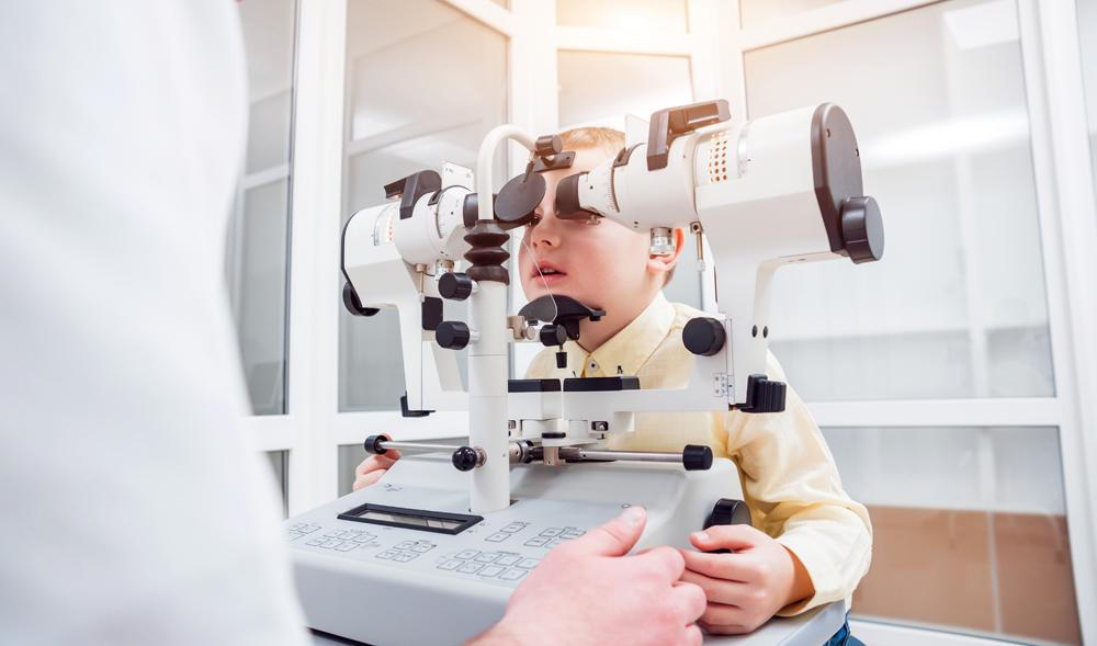
A crucial part in thorough ophthalmic evaluations, aesthetic area screening content plays a crucial duty in improving the analysis process for different eye conditions. By examining the full extent of a client's visual area, this examination supplies vital info about the functional integrity of the whole visual pathway, from the retina to the visual cortex.
Visual area testing is particularly important in the diagnosis and administration of conditions such as glaucoma, optic nerve disorders, and numerous neurological conditions that can influence vision. Via measurable dimensions of peripheral and central vision, medical professionals can detect refined modifications that might show the existence or development of these conditions, also prior to noticeable symptoms happen.
Moreover, visual area screening permits the tracking of treatment efficiency, helping eye doctors customize restorative treatments to private patients. eyecare near me. By tracking changes in visual area performance over time, doctor can make enlightened choices concerning adjusting medicines, recommending medical treatments, or executing other suitable actions to maintain or boost an individual's visual feature
Handling Macular Degeneration

Final Thought
Finally, progressed diagnostic tools play a critical duty in recognizing eye problems beforehand. Technologies such as Optical Comprehensibility Tomography and aesthetic area testing have significantly improved the precision and performance of detecting conditions like glaucoma and macular degeneration. Early detection enables prompt intervention and management of these problems, eventually causing better outcomes for people. It is essential for healthcare specialists to stay updated on these innovations to supply the finest possible take care of their individuals. eyecare near me.